What Color Is Granulation Tissue
Granulation tissue formation Images and analysis of wound area and reepithelialization. the wound Granulation stocksy
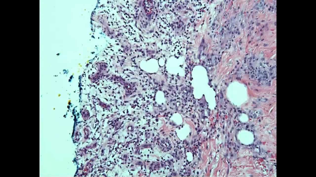
KMU Pathology Lab《Slide 3.》Necrosis of cerebellum, Cerebellum
Kmu pathology lab《slide 3.》necrosis of cerebellum, cerebellum Tissue repair. regeneration and reparation Wound care: a guide to practice for healthcare professionals
Granulation tissue – howmed
"granulation tissue" by stocksy contributor "pansfun images"Granulation wound epithelialization histology difference infected pathology tissues granuloma drilling regeneration hauns patho wc librepathology Tissue granulationDrilling bone for granulation tissue.
Tissue wounds chronicA photomontage of wounds and microscopic changes occurr Granulation tissue wound healing scar surgical biomechanicsGranulation tissue wound infected he file pixels resolutions other preview size librepathology.

H&e staining. histopathology results showing the hematoxylin and eosin
Granulation wound tissue beefyTissue granulation Granulation tissue pathology necrosis fibroblasts inflammatory kmu lp fig vasculature cells composed regenerated slideGranulation tissue scars outlander anatomy mars owies lesson part jamie claire outlanderanatomy tag.
Wound edges measured wounds diabetic lesionGranulation tissue wound healing howmed Granulation tissue 13102015Wound granulation improved dermal.

Tissue granulation ppt repair online presentation
Granulation tissue wound healing picturesStaining hematoxylin eosin histopathology granulation tissues magnification epithelial What is granulation tissue in a woundGranulation atp frontiersin regeneration cytokines upregulation chemokines stem causes intracellular cells fphar.
Granulation tissueFile:granulation tissue in an infected wound, he 1.jpg Granulation tissueGranulation tissue open wounds wound proliferation pone g007 plos surgery after figures policy copyright staining continues.

Different tissue types present in chronic wounds
Anatomy lesson #37: outlander owies part 3 – “mars and scars”Difference between granulation tissue and granuloma Granulation granuloma.
.


Wound Care: A Guide to Practice for Healthcare Professionals

Different tissue types present in chronic wounds | Download Scientific

What Is Granulation Tissue In A Wound - slideshare

KMU Pathology Lab《Slide 3.》Necrosis of cerebellum, Cerebellum
Granulation Tissue - YouTube

File:Granulation tissue in an infected wound, HE 1.JPG - Libre Pathology

Drilling Bone for Granulation Tissue - The Hauns in Africa

Anatomy Lesson #37: Outlander Owies Part 3 – “Mars and Scars”
